

1. Limb leads
Including standard limb leads I, II, and III and compression unipolar limb leads aVR, aVL, and aVF.
(1) Standard limb lead: also known as bipolar lead, which reflects the potential difference between the two limbs.
(2) Pressurized unipolar limb lead: in the two electrodes, only one electrode shows potential, and the potential of the other electrode is equal to zero. At this time, the amplitude of the waveform formed is small, so pressure is used to increase the measured potential for easy detection.
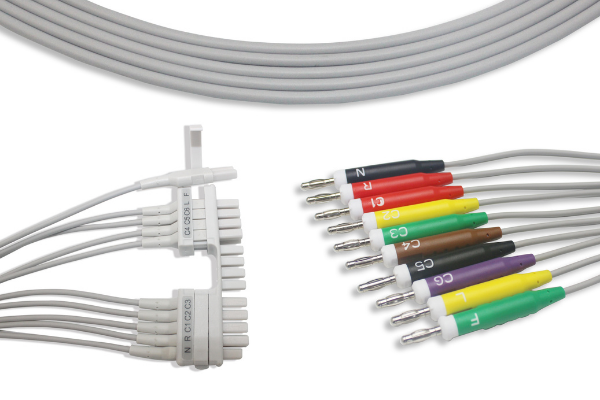
(3) When tracing the ECG clinically, there are 4 colors of the limb lead probe electrodes, and their placement positions are: the red electrode is on the wrist of the right upper limb, the yellow electrode is on the wrist of the left upper limb, and the green electrode is on the foot and ankle of the left lower limb. The black electrode is located at the ankle of the right lower limb.
2. Chest leads
It is a unipolar lead, including leads V1 to V6. During testing, the positive electrode should be placed on the specified part of the chest wall, and the 3 electrodes of the limb lead should be connected to the negative electrode through a 5 K resistor to form the central electrical terminal.
During routine ECG examination, 12 leads of bipolar, pressurized unipolar limb leads and V1~V6 can meet the needs. If dextrocardia, right ventricular hypertrophy, or myocardial infarction is suspected, lead V7, V8, V9, and V3R should be added. V7 is at the level of V4 at the left posterior axillary line; V8 is at the level of V4 at the left scapular line; V9 is at the side of the left spine Line V4 is at the level; V3R is at the corresponding part of V3 on the right chest.
Monitoring significance
1. The 12-lead monitoring system can reflect myocardial ischemia events in time. 70% to 90% of myocardial ischemia is detected by electrocardiogram, and clinically, it is often asymptomatic.
2. For patients at risk of myocardial ischemia, such as unstable angina and myocardial infarction, 12-lead ST-segment continuous ECG monitoring can promptly detect acute myocardial ischemia events, especially asymptomatic myocardial ischemia events, which is clinical Provide reliable basis for timely diagnosis and treatment.
3. It is difficult to accurately distinguish between ventricular tachycardia and supraventricular tachycardia with intraventricular differential conduction using only lead II. The best lead to correctly distinguish the two is V and MCL (the P wave and QRS complex have the clearest morphology).
4. When evaluating abnormal heart rhythms, using multiple leads is more accurate than using a single lead.
5. The 12-lead monitoring system is more accurate and timely to know whether the patient has arrhythmia than the traditional single-lead monitoring system, as well as the type of arrhythmia, onset rate, appearance time, duration, and changes before and after drug treatment.
6. Continuous 12-lead ECG monitoring is very important for determining the nature of arrhythmia, choosing diagnostic and treatment methods, and observing the effects of treatment.
7. The 12-lead monitoring system also has its limitations in clinical applications, and is susceptible to interference. When the patient’s body position changes or the electrodes are used for a period of time, a lot of interference waves will appear on the screen, which will affect the judgment and analysis of the electrocardiogram.
Post time: Oct-12-2021