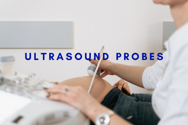
Ultrasonic probe (ultrasonic probe) is an indispensable key part of an ultrasonic diagnostic instrument. It can not only transform electric signals into ultrasound signals, but also transform ultrasound signals into electric signals, that is, it has the dual functions of ultrasound transmission and reception.
Classification of medical ultrasound probes
The structure and type of the ultrasound probe, as well as the conditions of the external excitation pulse parameters, work and focus mode, have a great relationship with the shape of the ultrasound beam it emits, and also have a great relationship with the performance, function, and quality of the ultrasound diagnostic apparatus. The transducer element material has little relationship with the shape of the ultrasound beam; however, the piezoelectric efficiency, sound pressure, sound intensity and imaging quality of its emission and reception are more related.
Pulse echo probe:
Single probe: It usually chooses piezoelectric ceramics ground into a flat thin disc as the transducer. Ultrasound focusing usually adopts two methods: thin shell spherical or bowl-shaped transducer active focusing and flat thin disc sound-dating lens focusing. Commonly used in A-type, M-type, mechanical fan scan and pulse Doppler ultrasonic diagnostic equipment.
Mechanical probe: The number of pressed electric chips and the movement mode can be divided into two types: unit transducer reciprocating swing scanning and multi-element transducer rotating switching scanning probe. According to the characteristics of the scan difference plane, it can be divided into sector scan, panoramic radial scan and rectangular plane linear scan probe.
Electronic probe: It adopts a multi-element structure and uses the principle of electronics to perform sound beam scanning. According to the structure and working principle, it can be divided into linear array, convex array and phased array probe.
Intraoperative probe: It is used to display the internal structure and the position of surgical instruments during the operation. It is a high-frequency probe with a frequency of about 7MHz. It has the characteristics of small size and high resolution. It has three types: mechanical scanning type, convex array type and wire control type.
Puncture probe: It passes through the corresponding body cavity, avoiding lung gas, gastrointestinal gas and bone tissue to get close to the deep tissue to be examined, improving the detectability and resolution. Currently there are transrectal probes,
Transurethral probe, transvaginal probe, transesophageal probe, gastroscopic probe and laparoscopic probe. These probes are mechanical, wire-controlled or convex array type; have different fan-shaped angles; single-plane type and multi-plane type. The frequency is relatively high, generally around 6MHz. In recent years, transvascular probes with a diameter less than 2mm and a frequency above 30MHz have also been developed.
Intracavitary probe: It passes through the corresponding body cavity, avoiding lung gas, gastrointestinal gas and bone tissue to get close to the deep tissues to be examined, improving the detectability and resolution. At present, there are transrectal probes, transurethral probes, transvaginal probes, transesophageal probes, gastroscopic probes and laparoscopic probes. These probes are mechanical, wire-controlled or convex array type; have different fan-shaped angles; single-plane type and multi-plane type. The frequency is relatively high, generally around 6MHz. In recent years, transvascular probes with a diameter less than 2mm and a frequency above 30MHz have also been developed.

Doppler probe
It mainly uses the Doppler effect to measure blood flow parameters, as well as the diagnosis of cardiovascular diseases, and can also be used for fetal monitoring. Mainly divided into the following three types:
1. Continuous wave Doppler probe: Most of the transmitter and receiver chips are separated. In order to make the continuous wave Doppler probe have high sensitivity, generally no absorption block is added. According to different uses, the way of separating the transmitting chip and the receiving chip of the continuous wave Doppler probe is also different.
2. Pulse wave Doppler probe: The structure is generally the same as the pulse echo probe, using a single-pressure wafer, with a matching layer and an absorption block.
3. Plum-shaped probe: Its structure is centered with only one transmitting chip, and six receiving chips around it, arranged in a plum blossom shape, used to check the fetus and obtain the fetal heart rate.
Post time: Nov-16-2021

